- Diabetes, Obesity and Metabolism
- Expression of LONP1 Is High in Visceral Adipose Tissue in Obesity, and Is Associated with Glucose and Lipid Metabolism
-
Ju Hee Lee, Saet-Byel Jung, Seong Eun Lee, Ji Eun Kim, Jung Tae Kim, Yea Eun Kang, Seul Gi Kang, Hyon-Seung Yi, Young Bok Ko, Ki Hwan Lee, Bon Jeong Ku, Minho Shong, Hyun Jin Kim
-
Endocrinol Metab. 2021;36(3):661-671. Published online June 22, 2021
-
DOI: https://doi.org/10.3803/EnM.2021.1023
-
-
4,519
View
-
151
Download
-
7
Web of Science
-
6
Crossref
-
 Abstract Abstract
 PDF PDF Supplementary Material Supplementary Material PubReader PubReader  ePub ePub
- Background
The nature and role of the mitochondrial stress response in adipose tissue in relation to obesity are not yet known. To determine whether the mitochondrial unfolded protein response (UPRmt) in adipose tissue is associated with obesity in humans and rodents.
Methods
Visceral adipose tissue (VAT) was obtained from 48 normoglycemic women who underwent surgery. Expression levels of mRNA and proteins were measured for mitochondrial chaperones, intrinsic proteases, and components of electron-transport chains. Furthermore, we systematically analyzed metabolic phenotypes with a large panel of isogenic BXD inbred mouse strains and Genotype-Tissue Expression (GTEx) data.
Results
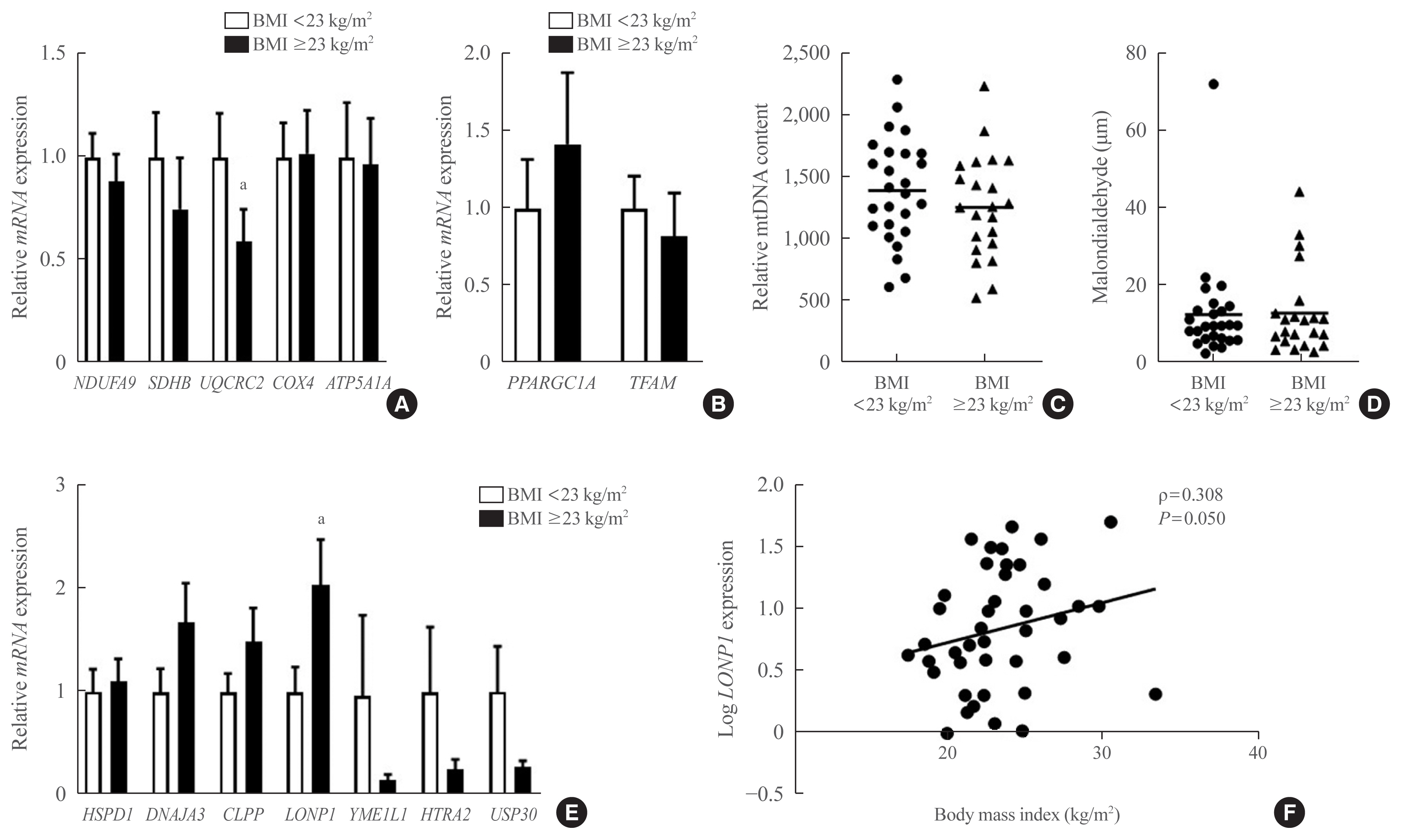
In VAT, expression of mitochondrial chaperones and intrinsic proteases localized in inner and outer mitochondrial membranes was not associated with body mass index (BMI), except for the Lon protease homolog, mitochondrial, and the corresponding gene LONP1, which showed high-level expression in the VAT of overweight or obese individuals. Expression of LONP1 in VAT positively correlated with BMI. Analysis of the GTEx database revealed that elevation of LONP1 expression is associated with enhancement of genes involved in glucose and lipid metabolism in VAT. Mice with higher Lonp1 expression in adipose tissue had better systemic glucose metabolism than mice with lower Lonp1 expression.
Conclusion
Expression of mitochondrial LONP1, which is involved in the mitochondrial quality control stress response, was elevated in the VAT of obese individuals. In a bioinformatics analysis, high LONP1 expression in VAT was associated with enhanced glucose and lipid metabolism.
-
Citations
Citations to this article as recorded by  - LONP1 ameliorates liver injury and improves gluconeogenesis dysfunction in acute-on-chronic liver failure
Muchen Wu, Jing Wu, Kai Liu, Minjie Jiang, Fang Xie, Xuehong Yin, Jushan Wu, Qinghua Meng
Chinese Medical Journal.2024; 137(2): 190. CrossRef - Mitochondrial quality control proteases and their modulation for cancer therapy
Jiangnan Zhang, Wenliang Qiao, Youfu Luo
Medicinal Research Reviews.2023; 43(2): 399. CrossRef - Effects of Obesity and Calorie Restriction on Cancer Development
Ekaterina Sergeeva, Tatiana Ruksha, Yulia Fefelova
International Journal of Molecular Sciences.2023; 24(11): 9601. CrossRef - Mitochondrial Dysfunction Associated with mtDNA in Metabolic Syndrome and Obesity
Natalia Todosenko, Olga Khaziakhmatova, Vladimir Malashchenko, Kristina Yurova, Maria Bograya, Maria Beletskaya, Maria Vulf, Natalia Gazatova, Larisa Litvinova
International Journal of Molecular Sciences.2023; 24(15): 12012. CrossRef - Down‐regulation of Lon protease 1 lysine crotonylation aggravates mitochondrial dysfunction in polycystic ovary syndrome
Yuan Xie, Shuwen Chen, Zaixin Guo, Ying Tian, Xinyu Hong, Penghui Feng, Qiu Xie, Qi Yu
MedComm.2023;[Epub] CrossRef - The mitochondrial unfolded protein response: A multitasking giant in the fight against human diseases
Zixin Zhou, Yumei Fan, Ruikai Zong, Ke Tan
Ageing Research Reviews.2022; 81: 101702. CrossRef
- A Case of Ectopic ACTH Syndrome Associated with Small Cell Lung Cancer Presented with Hypokalemia.
-
Hong Jun Yang, Hea Jung Sung, Ji Eun Kim, Hyo Jin Lee, Jin Min Park, Chan Kwon Park, Eun Suk Roh, Jae Hyung Cho, Seung Hyun Ko, Ki Ho Song, Yu Bai Ahn
-
J Korean Endocr Soc. 2007;22(5):359-364. Published online October 1, 2007
-
DOI: https://doi.org/10.3803/jkes.2007.22.5.359
-
-
1,958
View
-
26
Download
-
4
Crossref
-
 Abstract Abstract
 PDF PDF
- We report a case of a 73-year-old female patient who was diagnosed with ectopic ACTH syndrome caused by small cell lung cancer. We initially presumed that the patient was in a state of mineralocorticoid excess, because she had hypertension and hypokalemic alkalosis. This was however excluded because her plasma renin activity was not suppressed and her plasma aldosterone/plasma renin activity ratio was below 25. Moreover, her 24 hour urine free cortisol level was elevated and her serum cortisol levels after a low dose dexamethasone suppression test, were not suppressed. Furthermore, her basal plasma ACTH and serum cortisol levels increased and her serum cortisol level after a high dose dexamethasone suppression test was not suppressed. We performed studies to identify the source of ectopic ACTH syndrome and found a 3 cm-sized mass in the patient's right lower lobe of her lung, which was eventually diagnosed as small cell lung cancer following a bronchoscopic biopsy. In conclusion, Cushing's syndrome, and in particular ectopic ACTH syndrome, must be considered in the differential diagnosis of mineralocorticoid-induced hypertension. The excessive cortisol saturates the 11beta-hydroxysteroid dehydrogenase type 2 (11beta-HSD2) activity, which in turn, inactivates the conversion of cortisol to cortisone in the renal tubules. Moreover, excessive cortisol causes binding to the mineralocorticoid receptors, causing mineralocorticoid hypertension, characterized by severe hypercortisolism.
-
Citations
Citations to this article as recorded by  - Emergencia hipertensiva como debut de síndrome de Cushing paraneoplásico
E. Rubio González, M. de Valdenebro Recio, M.I. Galán Fernández
Hipertensión y Riesgo Vascular.2024; 41(2): 135. CrossRef - Management of small cell lung cancer complicated with paraneoplastic Cushing’s syndrome: a systematic literature review
Yanlong Li, Caiyu Li, Xiangjun Qi, Ling Yu, Lizhu Lin
Frontiers in Endocrinology.2023;[Epub] CrossRef - Ectopic Cushing Syndrome in Adenocarcinoma of the Lung: Case Report and Literature Review
Rana Al-Zakhari, Safa Aljammali, Basma Ataallah, Svetoslav Bardarov, Philip Otterbeck
Cureus.2021;[Epub] CrossRef - A Case of Ectopic Adrenocorticotropic Hormone Syndrome in Small Cell Lung Cancer
Chaiho Jeong, Jinhee Lee, Seongyul Ryu, Hwa Young Lee, Ah Young Shin, Ju Sang Kim, Joong Hyun Ahn, Hye Seon Kang
Tuberculosis and Respiratory Diseases.2015; 78(4): 436. CrossRef
- The Aging-related Change of Responses to TSH in Thyroid Cells.
-
Young Joo Park, Tae Yong Kim, Ji Eun Kim, Young Cheol Kim, In Kyeong Chung, Chan Soo Shin, Do Joon Park, Kyoung Soo Park, Seong Yeon Kim, Sang Chul Park, Hong Kyu Lee, Bo Youn Cho
-
J Korean Endocr Soc. 2004;19(2):141-151. Published online April 1, 2004
-
-
-
 Abstract Abstract
 PDF PDF
- BACKGROUND
To understand the mechanism of aging-related changes of the thyroid, the differentiated functions and growth of thyroid cells in response to TSH were investigated using aged or young thyrocytes. METHODS: FRTL-5 cells, with less than 10 or more than 45 passages, were used. After treatment with 1 U/L TSH or 1-100 mM NaI, the cAMP generation, iodide uptake, cellular proliferation or the expression of NIS mRNA or protein were measured. Sprague-Dawley rats were sacrificed at 5 and 16 weeks and 23 months, and their thyroids used for Northern blot analysis or immunohistochemistry of NIS. RESULTS: There were no differences in cAMP generation, iodide uptake, the proportions of G1/M or S phase, or intracellular DNA contents between the young and aged cells at basel levels. After TSH stimulation, these were increased in dose-dependent manners, with larger increments in the young cells. The changes in the NIS mRNA expression were similar in both the young and aged cells, but to a greater extent in the young cells. A similar phenomenon was observed in rat. However, the amount or intracellular distribution of NIS protein was not different. There was also no difference in the function or expression of NIS after treatment with a high dose of iodide. CONCLUSION: The aging-related decrease in the generation of cAMP might be thought of as one of the mechanisms of the decrement of iodide uptake or cellular proliferation with aging. The decreased expression of NIS mRNA seems to be the most important mechanism for the decreased iodide uptake capacity
- Small Medullary Thyroid Cancer Dectected by Genetic Mutation Screening in Men IIa Family.
-
Jae Hoon Chung, Kwang Won Kim, Ji Eun Kim, Byoung Joon Kim, Sung Hoon Kim, Kyung Ah Kim, Myung Sik Lee, Moon Gyu Lee
-
J Korean Endocr Soc. 1998;13(2):230-239. Published online January 1, 2001
-
-
-
 Abstract Abstract
 PDF PDF
- Multiple endocrine neoplasia (MEN) Ila is an inherited disease characterized by the development of medullary thyroid carcinoma, pheochromocytoma and hyperparathyroidism. It has been shown to be associated with germ-line mutatians in the RET proto-oncogene. Presymptomatic screening of medullary thyroid carcinoma in MEN IIa families enables the early diagnosis of this tumor with its significant morbidity, We describe a 19-year-old woman fmm a MEN IIa family who was founded by DNA analysis to be a gene carrier of MEN IIa and then was diagnosed, using a pentagastrin stimulation test, as having presymptomatie medullary thyroid carcinoma She underwent thyroidectomy and histologic examination confirmed medullary thyroid carcinoma. It is cancluded that direct genetic analysis for mutations in the RET proto-oncogene should be the diagnstlc test of choice for identifying family members at risk for MEN IIa and thyroidectomy on the basis of genetic analysis is a rational course of action.
|


